Multimodal AI for Biomedical Signal & Imaging Analysis
Modeling with image/waveform data and multi-omics integration for disease/target research, perturbation studies, clinical applications, and digital twin development.
Context
From Biomedical Data to Biological Understanding
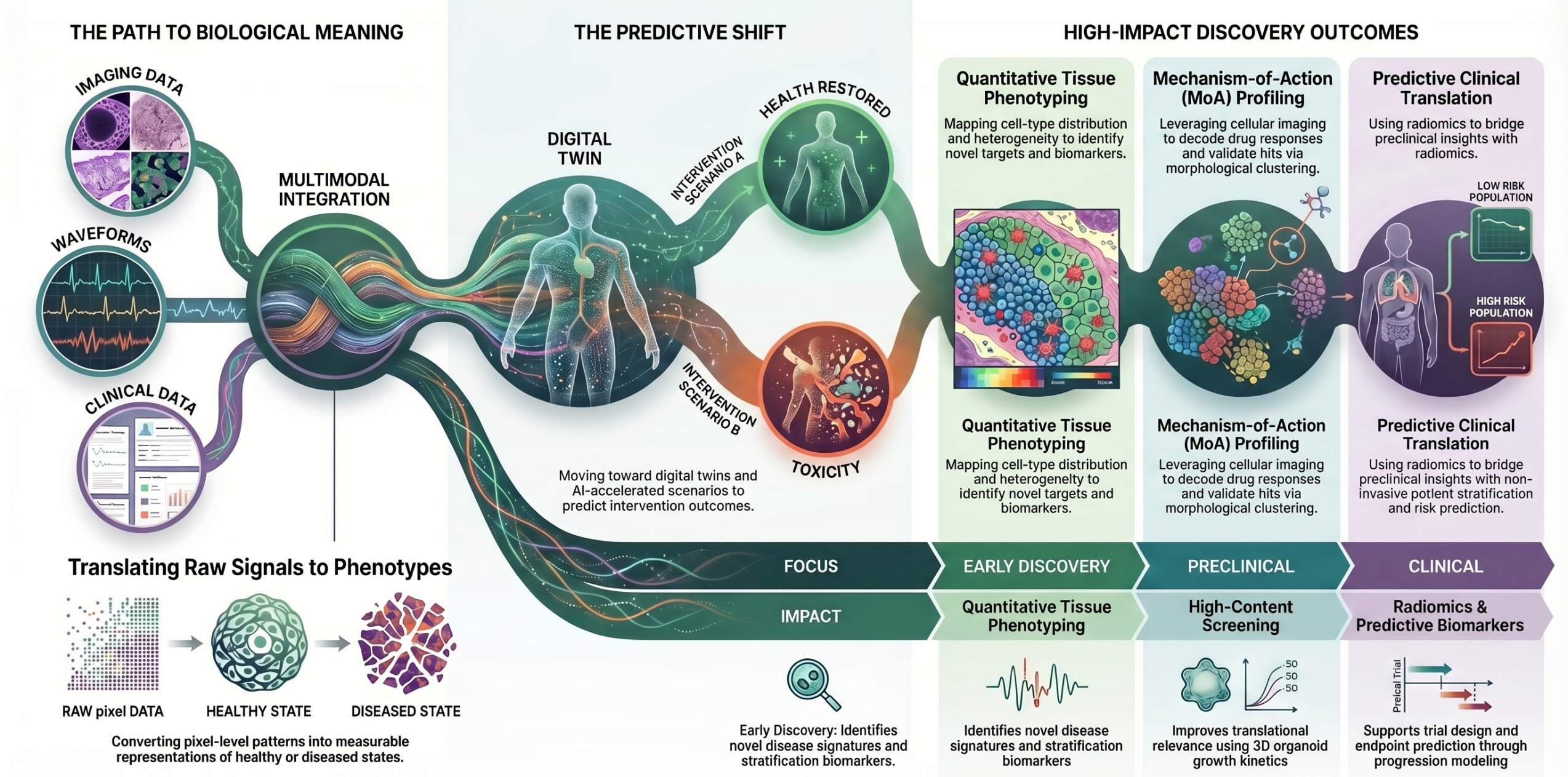
The goal of modern drug discovery is to understand physiology and derive actionable insights from biomedical data – spanning imaging, radiomics, and physiological waveforms. At its core, this comes down to answering a few key questions:
- What does healthy physiology look like?
- How does it change in disease?
- What happens when we intervene (gene perturbations or therapeutics)?
- Does the system return toward a healthy state, or diverge further?
- Are there early signals of toxicity or unintended effects?
Biomedical signals, such as imaging and waveforms, enable a seamless progression from understanding physiology → simulating response → guiding intervention.
Outcomes
Actionable Biological Insights Across the R&D Continuum
Quantitative Tissue Phenotyping for Target & Biomarker Discovery
Move beyond visual histopathology to high-resolution, quantitative tissue characterization.
- Cell-type detection, spatial distribution, and heterogeneity mapping
- Tumor grading and microenvironment profiling
- Extraction of morphology-derived biomarkers at scale
Impact:
Enables identification of novel targets, disease signatures, and stratification biomarkers directly from tissue architecture – supporting early-stage discovery and precision medicine strategies.
Mechanism-of-Action (MoA) and Phenotypic Profiling from Cellular Imaging
Leverage high-content imaging (e.g., cell painting) to decode cellular responses at scale.
- Phenotypic fingerprinting across perturbations (genetic or chemical)
- MoA prediction via morphological clustering
- Disease signature profiling and compound classification
Impact:
Accelerates hit validation and functional understanding, enabling researchers to connect compound activity to biological pathways without relying solely on target-based assays.
High-Content Screening and Efficacy Assessment in Complex Models
Quantify drug response in organoids, 3D cultures, and physiologically relevant systems.
- Growth kinetics, viability, and morphological response tracking
- Toxicity screening and phenotypic shifts
- Differentiation and structural organization analysis
Impact:
Improves translational relevance of preclinical studies, enabling more reliable assessment of efficacy and toxicity before clinical stages.
Predictive Imaging Biomarkers for Clinical Translation
Extract high-dimensional features from radiology and imaging modalities to enable non-invasive prediction.
- Radiomics-based feature extraction (shape, texture, intensity)
- Disease phenotyping and progression modeling
- Treatment response and risk prediction
Impact:
Supports patient stratification, trial design, and endpoint prediction, bridging preclinical insights with clinical decision-making.
Decode molecular signals directly from imaging data.
- Gene amplification and chromosomal abnormality detection
- Spatial gene expression mapping
- Integration with transcriptomics and multi-omics
Impact:
Enables spatially resolved biology, linking genotype to phenotype and unlocking insights not accessible through bulk omics alone.
Multimodal Integration: Imaging as a Core Layer in AI-Driven Biology
Integrate imaging with genomics, transcriptomics, and clinical data.
- Imaging + CRISPR perturbation analysis
- Imaging-driven reconstruction of gene networks
- Multimodal AI models for prediction and discovery
Impact:
Transforms imaging from a standalone modality into a central component of multi-scale biological modeling, enabling deeper mechanistic insights and improved predictive power.
Case Studies
Applied Case Studies
Myotube Fusion Analysis (Muscle Differentiation)
Quantitative analysis of skeletal muscle differentiation using iPSC-derived cells
- Key readouts:
- Myotube length
- Nuclei per muscle fiber
- Fusion index
- Enables comparison of wild-type vs diseased states
- Extends to 3D muscle models, capturing structural organization and maturation
Insight: Tracks how disease alters muscle formation and whether interventions restore normal differentiation
Adipocyte Phenotyping (Metabolic State Characterization)
Multi-channel imaging to capture cellular and metabolic state
- Key features analyzed:
- Lipid droplet accumulation (adipogenesis)
- Mitochondrial morphology (energy utilization)
- Cytoskeletal organization (cell shape changes)
- Cell boundary + nuclear quantification (size, proliferation)
Insight: Links morphological features to metabolic function, enabling precise characterization of adipocyte health and response
- ML-driven analysis of histopathology images (e.g., DMD vs healthy muscle)
- Detects:
- Fiber size variability
- Degeneration and regeneration markers
- Structural abnormalities (e.g., absence of tight junctions)
Insight: Differentiates healthy vs diseased tissue architecture, enabling objective and scalable pathology assessment.
Why Aganitha
The Aganitha Advantage
Biology-First AI
Our models are designed with domain expertise, trained and validated by scientists who understand the biological context, not just the pixel statistics.
2D to 3D Native
Native support for 2D and 3D imaging modalities, including organoid and tissue section analysis, without compromising on resolution or throughput.
Label-Free Intelligence
Brightfield and virtual staining pipelines reduce dependency on expensive fluorescent protocols while preserving analytical richness across phenotypic endpoints.
Multi-Modal Ready
Engineered for integration with genomics, transcriptomics, and proteomics, delivering a unified view of biological state that imaging alone cannot provide.